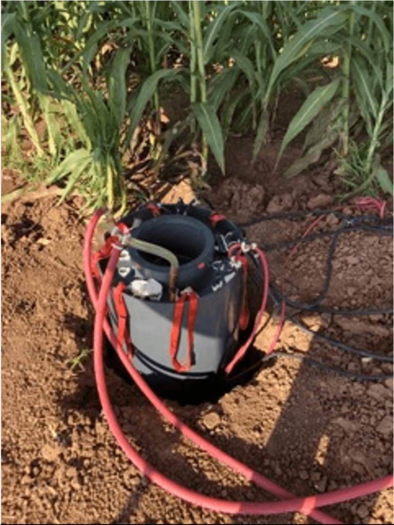
The architecture of plant roots influences many important functions, such as water and nutrient uptake. Root system architecture reflects the interaction of the genetic makeup of the plant with its environment. Characterizing this interaction (i.e., “root phenotyping”) provides critical information to plant breeders for developing new crop varieties that have improved drought tolerance, greater root biomass, and greater nutrient use efficiency. Phenotyping roots in the natural environment is important for understanding the impact of soil on how plants express their genetics. Imaging plant roots in undisturbed soil is very challenging, especially in soils with high clay content. A project that included SHI’s Chief Scientific Officer, Dr. Cristine Morgan, has been developing a field-scale device that can image these plant roots in natural soils – a mobile low-field Magnetic Resonance Imaging (LF-MRI) Rhizotron. This LF-MRI works on similar principles as the MRI machines used in human health. Figure 1 shows the LF-MRI in the field.
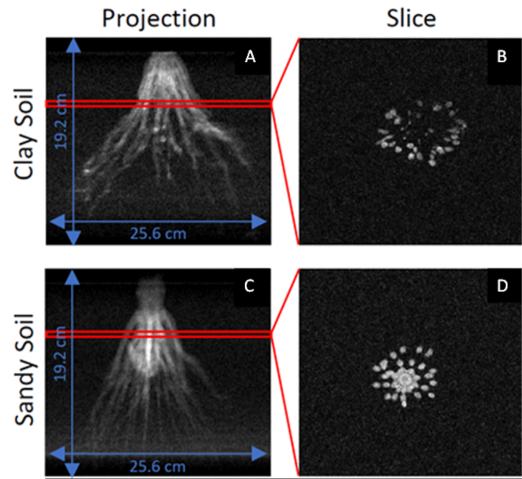
The project demonstrated that the LF-MRI Rhizotron works for root visualization and quantification. The research team from SHI, Texas A&M AgriLife Research, ABQMR Inc., Martinos Center for Biomedical Imaging, Harvard Medical School, and the National Institute of Standards and Technology found that the LF-MRI worked well for visualizing roots in moderate to high clay soils, demonstrating the potential for this technology; however, the broad application of this platform is a challenge because of the long scanning time needed to obtain 3D images. To address those challenges, the team is working to further improve the technology by increasing the magnetic field strength, resulting in faster scan times and higher resolution images. Figure 2 shows two root crown images produced by the new superconducting LF-MRI Rhizotron. Figure 2.A is an image of a sorghum root system that is 41 days old planted in a clay soil. Figure 2.B shows a slice of the image or a “bird’s eye” view of the root system. Figures 2.C and 2.D are corresponding images showing a sorghum root system of the same age grown in a sandy soil. This preliminary data is very promising, showing clear improvement in data quality and vast improvements in scanning time compared to the first system. This promising new technology for imaging undisturbed roots can open new insights about the structure of plant roots in soil and how plants and soil interact.
Read the publication here: http://dx.doi.org/10.1002/ppj2.20038
Phenotyping roots in the natural environment is important for understanding the impact of soil on how plants express their genetics, but imaging plant roots in natural conditions is very challenging, especially in soils with high clay content